Abdominal CT or ultrasound: what each scan shows and which one to choose
16 November 2026


16 November 2026
Your stomach hurts, the doctor orders a scan, and you wonder: why does one person get an ultrasound and another goes straight to CT? Can you play it safe and ask for a CT instead? And what if the ultrasound is normal but the pain does not go away?
Both tests look at the organs in your abdomen, but they work differently and answer different questions. Here is what each test covers, what it sees and misses, how it is done and how your doctor chooses between them.
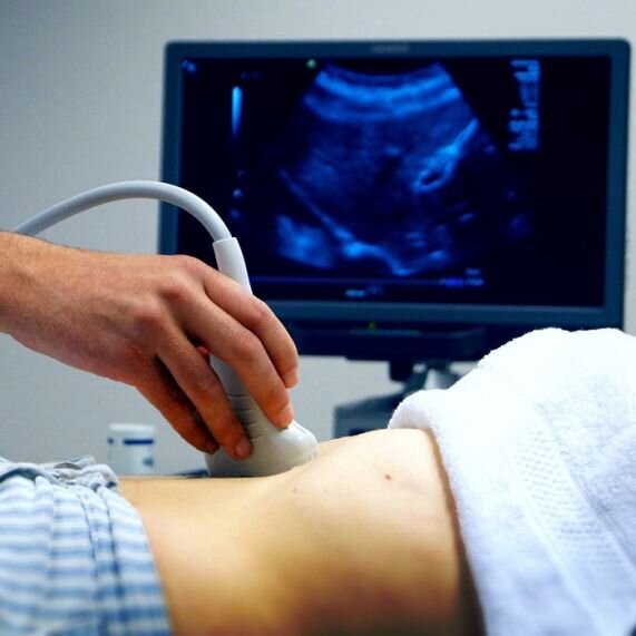
Ultrasound uses sound waves that bounce off your organs, and a computer turns them into a live image. There is no radiation. The MedlinePlus medical encyclopedia names this as the main advantage and says the test has no known risks.
A standard exam covers the liver, the gallbladder and bile ducts, the pancreas, the spleen and the large vessels: the portal vein and the abdominal aorta. The kidneys sit further back, behind the abdominal cavity, so many clinics, ours included, check them as a separate item or as part of a combined exam. Ultrasound shows the stomach and bowel only to a limited degree.
A CT scanner takes hundreds of thin X-ray slices from the top of the diaphragm down to the pelvis and builds them into a 3D image. The scan includes the liver, gallbladder, pancreas, spleen, stomach, bowel, kidneys, adrenal glands, blood vessels and lymph nodes, plus the lower ribs, spine and pelvic bones.
So a CT covers more ground than ultrasound. It shows the organs and everything between them: fluid, air, abscesses and enlarged lymph nodes. The abdomen is often scanned together with the pelvis, because many problems spread from one area to the other.
At Oberig Clinic, abdominal CT is done on Siemens Somatom go.Top and Somatom Confidence scanners with a reduced-dose mode, and ultrasound on an expert-class ACUSON Sequoia system by Siemens. You get your ultrasound report right after the exam, and radiologists in our Diagnostic Department read the CT scans.
Ultrasound sees soft tissue and fluid well. In practice it shows an enlarged liver, fatty liver, cysts, gallstones and polyps, a swollen pancreas in pancreatitis, an enlarged spleen and free fluid in the abdomen. MedlinePlus lists reasons for the test such as looking for gallstones and kidney stones, finding the cause of abnormal liver tests and checking a swollen organ.
It has known limits too. The patient guide RadiologyInfo explains that sound waves are disrupted by gas and air, so the bowel and organs behind it are harder to see. In larger patients, the extra tissue weakens the waves and the image gets less sharp. Sound waves cannot pass through bone at all.
There is also a human factor. It matters. The quality of an ultrasound depends on the doctor's experience and the machine. Two doctors may describe the same organ a little differently.
CT is not bothered by bowel gas or extra weight, and it sees bones and everything behind them. MedlinePlus lists what an abdominal CT can find: masses and tumours, including pancreatic cancer and lymphoma, infections and injuries, kidney stones, appendicitis, abscesses, pancreatitis and its complications such as pancreatic abscesses and pseudocysts.
That makes CT especially useful for sudden severe belly pain, after injuries, for finding and staging tumours, and when complications are suspected. The price of this accuracy is radiation and, often, a contrast injection.
| Ultrasound | CT | |
|---|---|---|
| How it works | Sound waves | X-rays, hundreds of slices |
| Radiation | None | Yes, low |
| Time | Usually under 30 minutes | Scan takes seconds, visit about 15 minutes |
| Bowel gas, extra weight | Make the image worse | Hardly matter |
| Sees best | Liver, gallbladder, fluid, cysts | Tumours, abscesses, injuries, stones in the ureter, lymph nodes |
| Repeat | As often as needed, also for children and in pregnancy | Only when medically needed |
| Contrast | Not needed | Often needed |
This table sums up information from MedlinePlus, RadiologyInfo and our clinic pages. Your doctor, who knows your symptoms, always makes the final choice.
You lie on a couch, the doctor puts gel on your belly and moves a probe over it, sometimes asking you to hold your breath or turn on your side. MedlinePlus notes that the test usually takes less than 30 minutes. No pain, no needles, and you get the result straight away. You do need to prepare: the exam is done on an empty stomach so the gallbladder is full and there is less gas in the bowel.
You lie on a table that moves through the ring of the scanner and hold your breath when asked. The scan itself takes seconds. If contrast is needed, it goes into a vein, and sometimes you drink it beforehand. According to MedlinePlus, with IV contrast you may be asked not to eat or drink for 4 to 6 hours before the test. At our clinic, before an abdominal CT you drink up to 1 litre of plain water in portions over 1 to 1.5 hours.
Doctors usually start with ultrasound. It is easy to get, safe and answers most routine questions about the liver, gallbladder, pancreas and spleen. You go straight to CT when:
It works the other way too. Sometimes ultrasound wins. It shows small gallstones and polyps better than CT, and doctors prefer it for pregnant women and children to avoid radiation.
Without contrast, CT shows kidney and ureter stones, free air from a perforation, calcifications and bones well. Contrast makes vessels, tumours and inflamed areas stand out, so it is used more often for cancer questions and to check the blood supply to organs. Your doctor decides if you need it, and kidney function is often checked first. We cover contrast safety in a separate article.
For a detailed look at the liver, spleen and pancreas, there is a dedicated CT of the liver, spleen, pancreas and stomach with a targeted protocol.
Not without a reason. Here is why. CT means radiation and often contrast, while ultrasound answers many questions just as well. Your doctor picks the test for the specific question.
Do not stop there. Show the result to your doctor. Depending on your symptoms, you may need blood tests, a gastroscopy, a colonoscopy or a CT. A normal ultrasound does not rule out stomach and bowel problems, which ultrasound sees poorly.
Ultrasound is usually enough for a first look. CT is needed to clarify a growth that was found, check the blood vessels or see how far a tumour has spread.
Yes, you can do both in one visit. Staff will tell you the order and how to prepare for each when you book.
Yes, it is usually done on an empty stomach. Staff will give you the exact instructions when you book.
Most often a gastroenterologist, a surgeon, a urologist or an oncologist. You can also book on your own, but it is best to go over the results with a doctor.
If you have belly pain or your doctor has ordered an abdominal scan, you can have both tests in one place at Oberig Clinic. Abdominal ultrasound is done on an expert-class machine with a report on the spot, and abdominal CT on Siemens scanners, read by radiologists in our Diagnostic Department. If needed, a gastroenterologist or surgeon at the clinic will explain the results.
Book by phone at +38 044 521 30 03 or online.