CT vs MRI: what is the difference and which one suits you
9 November 2026


9 November 2026
Your doctor says "get a scan", and the question comes right away: which one? Both CT and MRI produce cross-sectional images of your organs, and both look like a big machine with a table. Are they the same thing? No. They work differently and see different things. The right choice always depends on what exactly needs to be found.
Below we explain in plain words how both scans work, what suits the head, spine, belly, pelvis and joints and cancer care best, what's safer and what costs more. Most importantly, why your doctor, not you, makes the final call.
Computed tomography is an X-ray, just a very precise one. A tube rotates around your body and takes hundreds of thin slices, and a computer assembles them into a 3D image. According to the NHS, CT is used to check bones and internal organs after an injury, find the cause of symptoms, see how well treatment is working and even guide procedures such as biopsies.
Magnetic resonance imaging works without X-rays. The machine creates a strong magnetic field and radio waves, and a computer "listens" to how tissues respond and builds an image. The MSD Manual stresses that unlike CT and PET scans, MRI doesn't use ionizing radiation.
What's the difference for you as a patient? A CT takes a few minutes, and the scan itself only seconds. An MRI lasts 20 to 60 minutes, the machine is loud and you have to lie still. That can be hard if you're claustrophobic. Very hard for some. On the other hand, MRI involves no radiation, so it can be repeated as often as needed to track treatment.
| Feature | CT | MRI |
|---|---|---|
| How it works | X-rays | Magnetic field and radio waves |
| Radiation | Yes, dose depends on the area | None |
| Duration | Minutes, scanning takes seconds | 20-60 minutes |
| Best at showing | Bones, lungs, fresh bleeding, emergencies | Soft tissue: brain, spinal cord, discs, ligaments, pelvic organs |
| Metal in the body | No problem | Pacemakers and some implants may rule it out |
| Contrast | Iodine-based | Gadolinium-based |
| Cost | Usually lower | Usually higher |
In short, the two differ in how they work, how long they take, radiation and which tissues they show best.
Neither is more accurate "in general". CT wins where dense structures and speed matter: fractures, lungs, bleeding, stones, an acute belly. MRI wins where similar soft tissues need to be told apart: the gray and white matter of the brain, spinal discs and nerves, cartilage and ligaments, layers of the bowel wall, pelvic organs.
The question isn't what's better overall. It's what's better for a specific task. Here are the most common situations.
| Area or task | Usual choice | Why |
|---|---|---|
| Head, suspected stroke in the first hours | CT | Quickly shows bleeding, which shapes treatment |
| Brain: tumors, multiple sclerosis, epilepsy | MRI | Best view of brain tissue and small lesions |
| Spine: herniated discs, nerve roots, spinal cord | MRI | Shows discs, nerves and the spinal cord |
| Spine: fractures, bone changes | CT | Shows bone in detail |
| Lungs | CT | Air and lung tissue contrast sharply on X-ray |
| Abdomen, emergencies | CT | Fast, shows bleeding, perforation, stones |
| Liver, pancreas, bile ducts | MRI or CT | MRI characterizes lesions and ducts more precisely |
| Pelvis: uterus, ovaries, prostate, rectum | MRI | Best at telling tissue layers apart |
| Knee and other joints | MRI | Shows menisci, ligaments, cartilage |
In the first hours of a stroke or after a head injury, CT has the edge: it shows within seconds if there's bleeding. That decides if clot-dissolving drugs can be given. We describe this scan in detail on our brain CT scan page. For planned checkups and looking for tumors, multiple sclerosis lesions or causes of epilepsy, a brain MRI is better. For the head, it's not about quality but about the task: an emergency or a detailed look.
For back pain spreading down the leg, a suspected herniated disc or pressure on nerve roots or the spinal cord, MRI is the choice. CT helps with injuries, fractures, bone changes and when MRI isn't possible, for example because of a pacemaker.
For sudden belly pain, injury, suspected bleeding or perforation, CT is faster and more available. For a detailed look at lesions in the liver, pancreas or bile ducts, MRI is often chosen. It's better at telling benign from malignant growths. For the bowel, it depends: CT for emergencies, MRI for Crohn's disease and rectal tumors, and colonoscopy for the most accurate view of the lining.
What about the pelvis? MRI, as a rule: it shows the uterus, ovaries, prostate and rectum in detail. The knee? Also MRI, because menisci, ligaments and cartilage are hard to see on CT.
For the lungs, CT has almost no rivals. It finds small nodules, inflammation, signs of fibrosis and, with contrast, blood clots in the lungs. Modern machines can image the lungs with MRI, but it's used only in narrow cases, for example in children or pregnancy, when avoiding radiation matters.
For the heart and vessels, each scan has its job. CT coronary angiography shows narrowed heart arteries and calcium in them. Cardiac MRI assesses the heart muscle, scars after a heart attack, inflammation and congenital defects. For neck and brain vessels, CT or MR angiography is used, depending on how urgently an answer is needed and whether there are contraindications.
Doctors try to avoid radiation in children, so when there's a choice, ultrasound or MRI comes first. But it's hard for a small child to lie still for half an hour, and MRI is sometimes done under sedation. CT takes seconds. It's used in children after injuries or in emergencies when the result affects treatment.
After a fall or blow, when a fracture needs ruling out fast, an X-ray comes first, and CT follows for complex fractures and injuries to the pelvis or spine. It shows bone fragments and displacement in 3D, and that helps the surgeon plan. When a knee, shoulder or hip hurts without an obvious fracture, MRI shows damage to menisci, ligaments, tendons and cartilage that CT can't.
In cancer care, the two scans complement each other. CT of the chest and abdomen is the standard for finding spread and tracking treatment. MRI is more precise for tumors of the brain, liver, pelvis, rectum and breast. Patients often have both at different stages, and the oncologist sets the plan. To track treatment, repeat scans should use the same method so results can be compared.
The key difference is radiation. CT uses X-rays, and although modern machines cut the dose considerably, CT isn't done without a reason, especially in children and pregnancy. MRI uses no radiation, and on that front the answer is clear. But MRI has its own limits: pacemakers, some metal implants and fragments, and severe claustrophobia.
Both may need contrast, and each has its own safety rules. We cover radiation doses, contrast and implants in a separate article.
For a CT, you lie on a table that slowly slides through a wide ring. The radiographer will ask you to hold your breath for a few seconds so the images stay sharp. If contrast is needed, it's injected into a vein, and you may feel a brief warm flush. The whole thing takes a few minutes.
For an MRI, you lie inside the machine's tunnel. A special coil is placed around the area being scanned. The machine knocks loudly, and you'll get headphones or earplugs. You hold a call button, and the staff can see and hear you the whole time. Your main job is to stay still. A head scan takes 20-30 minutes, the abdomen and pelvis 30-45 minutes, rarely up to an hour. Neither scan hurts. The only unpleasant parts are the MRI noise and lying still, plus a needle in your vein if contrast is used.
If you're afraid of enclosed spaces, mention it in advance. Modern scanners are wider than older ones, and your doctor can offer something to help you relax.
Contrast makes blood vessels and some tissues brighter on the images. It helps tell a tumor from healthy tissue, spot inflammation and assess blood flow. CT uses iodine-based agents, MRI uses gadolinium-based ones. Your doctor decides if contrast is needed and checks for allergies and kidney function first.
The best approach is simple: come with a specific question from your doctor and your previous results. Then the radiologist can tailor the scan to your situation, and the report will be truly useful.
After a CT or MRI, you receive a written description, the radiologist's conclusion and the images on a disc or via a link. The report isn't a diagnosis. The doctor who referred you makes the diagnosis by matching the images with your symptoms, exam and tests. At Oberig clinic, brain MRI scans get two independent reviews before the report is issued.
Your doctor decides on the scan. They know what they're looking for and choose the method that answers the question best. A scan you pick yourself because it seems "more expensive and better" often misses the needed information, and the test has to be repeated. That costs time and money. If you've been referred for one and have doubts, ask your doctor why.
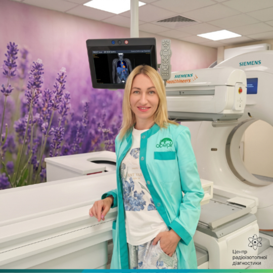
Oberig clinic offers both. Magnetic resonance imaging is done on Siemens Magnetom Vida 3 Tesla and Magnetom Sempra 1.5 Tesla scanners, and computed tomography on Siemens Somatom go.Top and Somatom Confidence machines with reduced radiation doses. The radiologist can suggest the best method for your case and, if needed, do both scans in one visit without you traveling between centers.
Usually CT. The price depends on the area and contrast, but MRI generally costs more. Current prices are on our CT and MRI pages.
Often yes, if your doctor thinks it's useful. If both scans use contrast, the doctor will check your kidney function and the gap between injections.
MRI without contrast can be repeated as often as medically needed. CT is done when the benefit outweighs the radiation risk, and your doctor decides how often. More in our separate safety article.
You can, but it's rarely a good idea. Without a clear medical question, it's easy to pick a scan that won't show the problem. Talk to a doctor first, even briefly. It saves money. And time.
No. In many situations X-ray or ultrasound remains the first step, and a scan is ordered to clarify things.
MRI without contrast can be done if needed, especially after the first trimester. CT is done in pregnancy only when it's vital.
It combines CT with a test that shows how active tissues are. It's used in cancer care to find active tumor sites. It's a separate method, ordered by an oncologist.
If your doctor has ordered a scan and you're not sure which one fits best, give us a call. Oberig clinic's radiologists will tell you what method will answer your question and book you at a convenient time. Scans are done on Siemens equipment, and radiologists write the reports.
Book by phone at +38 044 521 30 03 or online.